
Dr. Daniela Popescu, Professor in the Department of Biological Sciences at Kent State University’s Geauga campus, has earned a reputation for incorporating nontraditional teaching techniques into her courses. From her use of interactive i>clickers in the classroom to museum field trips and involvement in new research projects, she employs an ever-evolving use of art and technology to drive home human anatomy and physiology lessons.
Most recently, Dr. Popescu received a Teaching Development Grant to purchase a 3D printer for the Geauga campus. She was investigating the potential benefits of incorporating 3D‑printing activities into her Anatomy and Physiology laboratory courses.
“I always try to identify nontraditional teaching tools and learning approaches that I would have loved to have had access to when I was a student, whether I am visiting art or science museums or attending a conference,” she explains.
EXPECTATIONS
Dr. Popescu envisioned using the 3D printer to create various human anatomy models, designing model-based activities to enhance students’ learning experiences. She anticipated that using these three-dimensional models would improve students’ academic performance, motivation, collaboration, active participation, and communication in class.
She first learned about the use of 3D‑printed models in A&P courses while attending the 2017 Human Anatomy and Physiology Society Annual Conference in Salt Lake City.
“The idea stayed with me ever since,” says Dr. Popescu. “I was very happy and grateful when I was finally able to implement this idea with the gracious support of the University Teaching Council and our Dean, Dr. Angela Spalsbury, who is always very supportive and encourages the incorporation of innovative methods of teaching.”
With this support and the assistance of Kent State Geauga’s IT Manager Kevin Hilger, a Dremel 3D40-FLEX EDU printer was acquired and began to be used in the A&P laboratory courses by Fall Semester 2024.
Neither Prof. Popescu nor Hilger had prior hands‑on experience with 3D printing, so they completed Dremel’s online training courses to learn how to use the modeling software to create 3D-print models of human bones and other organs.
“Once we began producing anatomy models for her class, we quickly discovered that some prints required more than six hours to complete,” says Hilger. “To keep the process moving, our IT student worker, Nathan Vulinec, often started print jobs first thing in the morning whenever Professor Popescu needed a new model.”
Initially, Dr. Popescu designed an activity that focused on the skeletal system, one of the most challenging organ systems to learn, due to its multiple components and complexities.
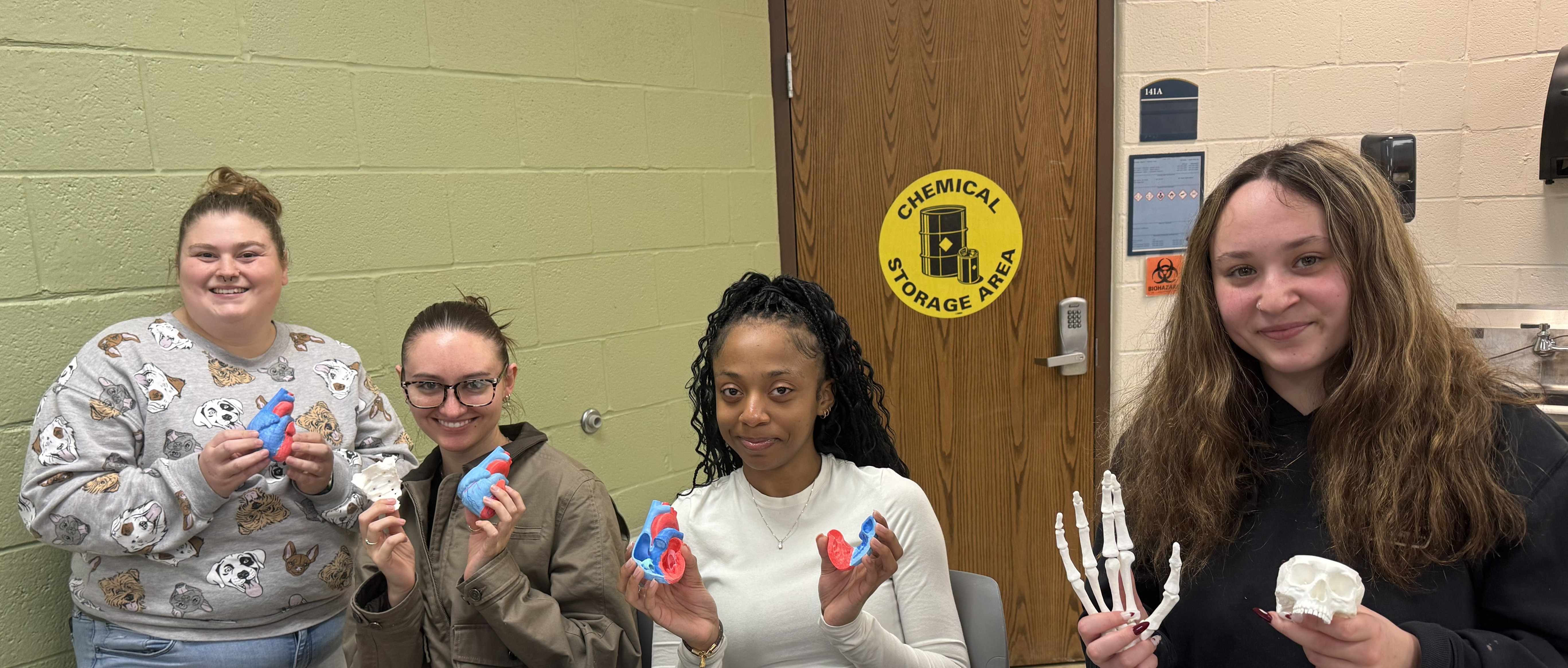
At the start of the Fall 2024 semester, students selected one or more bones of interest from a list, then took the pre-3D-printed bone models home as reference material to write an essay on the anatomy of their chosen bone(s) and to prepare a team presentation to the class.
Dr. Popescu has also incorporated 3D printing into her A&P II course involving the cardiovascular system. Students are organized into teams, each of which is responsible for painting a white 3D-printed heart model, including the blood vessels associated with each chamber of the heart. Students can use either blue or red colors, with blue indicating blood vessels that carry deoxygenated blood, and red for blood vessels that carry oxygenated blood.
The multi-sensory activity encourages students to manipulate the 3D models, collaborate together, interact with their professor, and use kinesthetic and visual learning modalities to help them master the anatomy of the heart and the characteristics of the blood vessels associated with each chamber of the heart.
OUTCOMES
Hilger shares, “Having the ability to print anatomy models on demand has been a major advantage for student learning. Traditional models are limited in quantity, but the 3D printer allows us to create additional resources whenever needed, giving students greater access to hands‑on materials. It also opens the door to hands‑on class projects, allowing students to mark or modify low‑cost printed models in ways that wouldn’t be possible with standard commercial sets.”
After completing their projects, students were surveyed about their use of the 3D-printed models in their coursework. “The positive feedback I received from students encouraged and motivated me to continue using the 3D-printed anatomy models in our A&P courses,” says Dr. Popescu.
“I believe that this hands-on approach using the 3D-printed human anatomy models enables our students to develop a deeper understanding of the bones, bone markings, and the relationship of bones to one another, as well as a deeper understanding of the anatomy of the heart and the blood vessels associated with each chamber of the heart.”
In fact, Dr. Popescu observed that her A&P II students performed significantly better on their Laboratory Test 1 in 2025 compared with 2024, before the 3D-printing activities were incorporated into the curriculum.

Dr. Popescu will not only continue using the 3D-printed models in her A&P laboratory courses, but also looks forward to designing other exciting 3D-printing activities for her students in the future.
Student Feedback
Ellori Harsch and Kelsey Boyd are two students who used the 3D-printed models of human bones and hearts in Dr. Popescu's Anatomy & Physiology I & II classes. They volunteered to share their experiences using the tridimensional learning tools.
ELLORI HARSCH
An Akronite, Ellori is majoring in the Bachelor of Science in Nursing program, anticipating graduation in 2029.
“With these models, not only were we able to physically understand the anatomical structure—as the models are very detailed—having these physical models further consolidated our understanding and knowledge of these body parts, far more than any diagram would,” Ellori says.
“When learning about the different human bones and organs, their physiological processes, and their anatomical significance, it can be difficult to imagine these structures in their entirety. Yes, we have access to models in the lab, but being able to have a model with you and study it throughout the weeks really added to the interactive learning experience. The 3D printer adds a new level of interactive teaching and learning that further encourages students' memory of detail and understanding of a subject.”
Ellori notes, “Being at a regional campus, I’m very thankful we have access to such incredible technology, and such incredible professors!”
KELSEY BOYD
Kelsey grew up in the small farming community of Gustavus, Ohio. She is a guest student at Kent State, taking post-baccalaureate classes in preparation for a Physician Assistant program.
“The models allowed us to examine the shapes, spatial relationships, and details of the heart and bones in a way that images alone cannot fully show,” Kelsey shares. “Painting the heart to follow the paths of oxygenated versus deoxygenated blood really helped drive home key concepts by turning abstract diagrams into tangible learning tools.”
Using 3D printing made the information they were learning feel more concrete and engaging, she reports. “Handling the models helped me visualize the three-dimensional arrangement of structures in the body, which is difficult to grasp from flat images. It also encouraged active and group learning because we were not just memorizing parts but physically exploring how they fit together and function. This hands-on experience deepened my understanding of anatomical relationships and physiological processes, making the material easier to remember and apply.”
Dr. Popescu's interactive, hands-on approach to learning also made the classes more engaging and effective for Kelsey. “Instead of relying solely on lectures, the use of activities based on 3D-printed structures allowed students to actively participate in the learning process, encouraging curiosity, discussion, and collaboration while helping students better understand complex concepts. Overall, it made the subject matter more memorable, and it helped connect theoretical knowledge with practical experience.”